Shape Meets Function
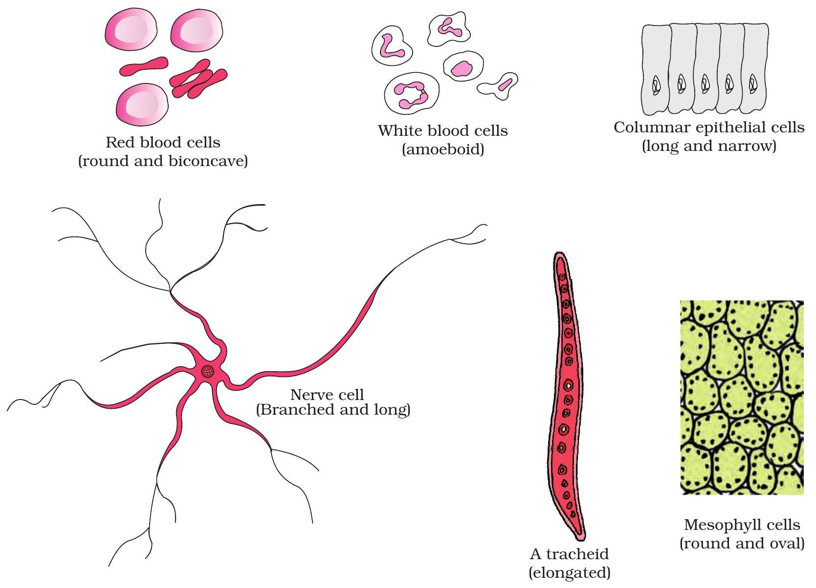
Red blood cell, neuron and tracheid—contrasting geometries, common purpose: efficiency.
Form follows function
Biconcave red blood cells squeeze through tiny capillaries, exposing maximum surface for rapid gas exchange.
Branching neurons speed impulses across metres, while narrow, lignified tracheids channel water upward; each shape serves its task.
Key Points:
- Disc-like RBC → high surface-area-to-volume ratio, flexible flow.
- Tree-like neuron → wide reach for rapid, directed signalling.
- Tube-like tracheid → capillary pull of water and structural support.
- Challenge: How could sickled RBCs reduce oxygen delivery?
Sizing Up Cells
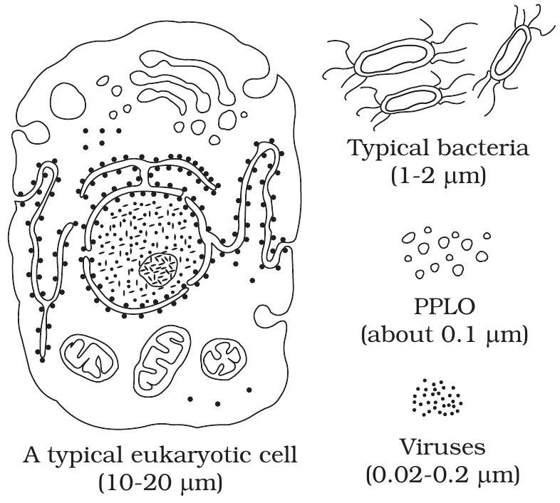
Scale bar compares virus, bacterium and eukaryotic cell.
From Nanometres to Micrometres
Viruses average 100 nm, relying on host cells because they hold few molecules.
Prokaryotes, about 1 µm, divide swiftly; diffusion easily reaches every corner.
Eukaryotes grow 10–100 µm; their low surface-area/volume ratio demands organelles for transport and energy.
Key Points:
- Virus < Prokaryote < Eukaryote in size: ~0.1 µm → 1 µm → 20 µm+
- Complexity rises with size; organelles solve transport and energy limits.
- Typical bacteria are too small for mitochondria—insufficient room and surplus surface already meets energy needs.
Cellular Power Plants
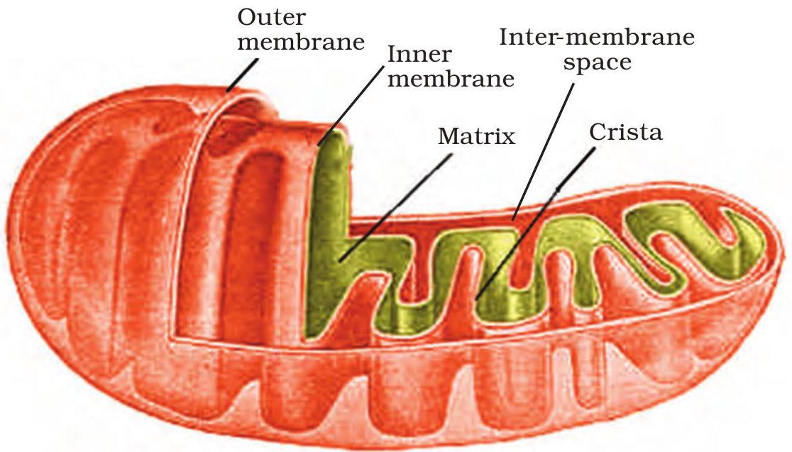
Mitochondrion (left) and chloroplast (right)
Same blueprint, different fuels
Cristae and thylakoids are folded or stacked membranes that multiply reaction surface, revealing the organelles’ shared design logic.
On cristae, electron transport drives ATP formation; on thylakoids, light energy powers glucose assembly, later yielding ATP.
Key Points:
- Cristae: inward folds pack electron-transport chains for rapid ATP output.
- Thylakoids: stacked discs (grana) spread chlorophyll to capture photons efficiently.
- Mitochondria convert food to ATP directly; chloroplasts store energy first as glucose.
- Both retain circular DNA & ribosomes — strong evidence for an endosymbiotic origin.
Motility Structures 9+2

Decode the 9+2 Axoneme
Cilia and flagella share a 9+2 array—nine peripheral microtubule doublets surrounding two central singlets.
Each axoneme sprouts from a basal body, a modified centriole anchoring the structure under the plasma membrane.
Key Points:
- 9+2 axoneme = 9 doublets + 2 singlet microtubules.
- Basal body templates and anchors each motile appendage.
- Dynein-driven sliding bends the axoneme; mutations cause immotile cilia and chronic respiratory disease.