Cells Come in Many Shapes
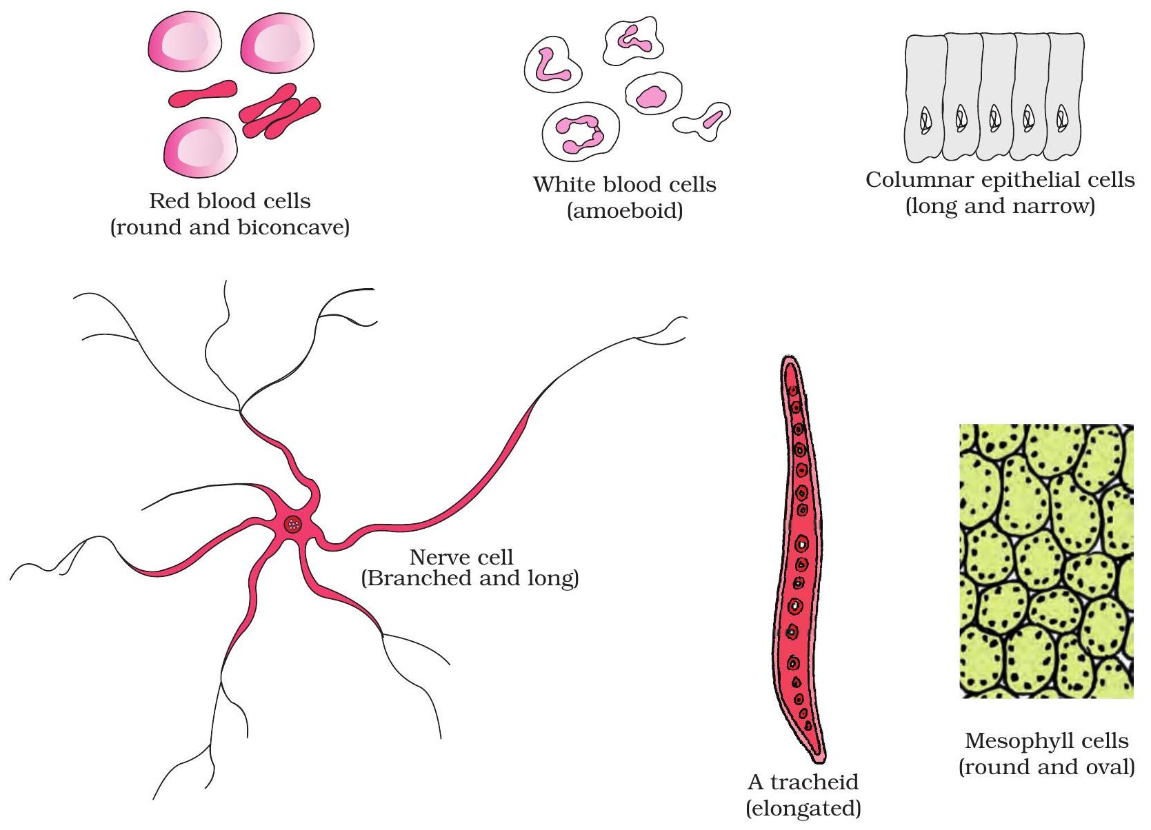
Diagram: RBC, nerve, columnar epithelium and WBC
Shape supports function
Cells vary widely. Each outline equips the cell for its task.
Spotting these forms lets us link structure to role.
Key Points:
- RBC – round, biconcave; squeezes through narrow capillaries.
- Nerve cell – long with branches; carries impulses over distance.
- Columnar cell – tall pillar; absorbs and protects linings.
- WBC – irregular, amoeboid; slips out to attack microbes.
Drag each label to its matching cell on the picture.
Plant vs Animal Cell
Shared & Unique Parts
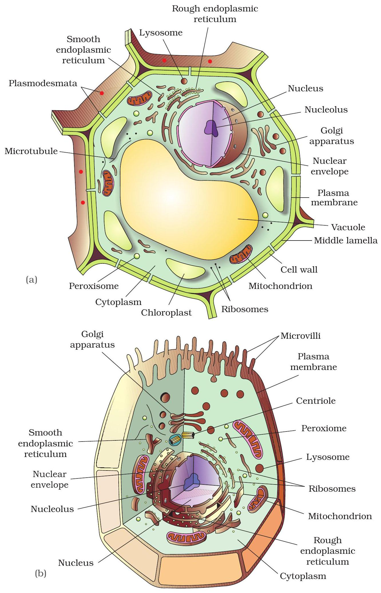
Both plant and animal cells contain nucleus, endoplasmic reticulum, Golgi bodies and mitochondria.
Plant cells add a rigid cell wall, large central vacuole and chloroplasts. Animal cells add centrioles and small temporary vacuoles.
Tap the chloroplast and the centriole in the diagram to test your understanding.
Key Points:
- Common: nucleus, ER, Golgi, mitochondria
- Plant-only: cell wall, chloroplast, large vacuole
- Animal-only: centriole, small vacuoles
Mitochondria: Powerhouse
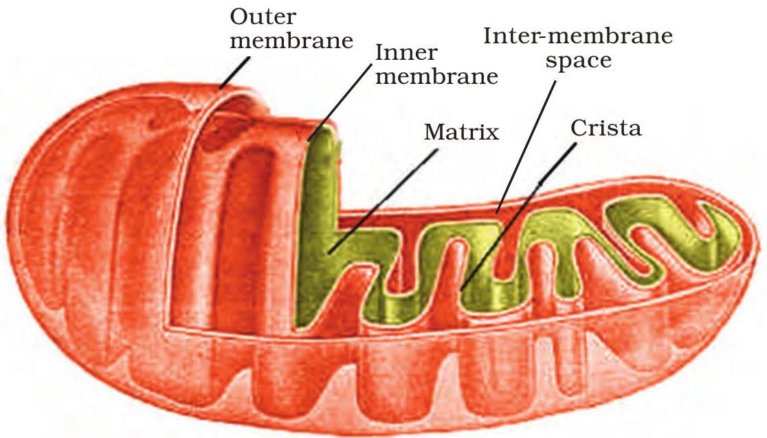
Folded cristae visible inside the double membrane.
Cristae & Matrix Fuel ATP
Mitochondrion has an outer membrane and a deeply folded inner membrane called cristae.
Cristae greatly enlarge the surface that holds the electron transport chain and ATP synthase.
The central matrix contains Krebs-cycle enzymes, circular DNA and 70 S ribosomes, allowing some protein synthesis.
Reactions on cristae and in the matrix together generate most cellular ATP.
Key Points:
- Folded cristae = larger surface → more ATP output.
- ATP synthase sits on cristae membranes.
- Matrix enzymes drive Krebs cycle and contain bacterial-like DNA & 70 S ribosomes.
Chloroplast: Green Factory
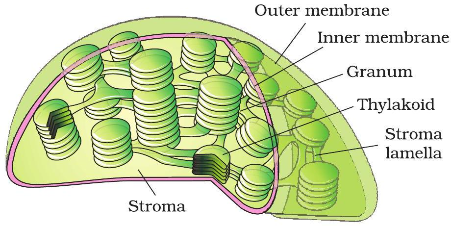
Stacks of grana inside a chloroplast
How the Parts Work Together
Chloroplasts contain flattened sacs called thylakoids, stacked into grana.
Thylakoid membranes hold chlorophyll that captures light and forms ATP + NADPH.
Energy moves into stroma, where enzymes fix CO₂ and build glucose.
Key Points:
- Light-dependent reactions occur on thylakoid membranes.
- Calvin cycle enzymes in stroma use ATP and NADPH.
- Result: sunlight + CO₂ → energy-rich sugars.
ER ➔ Golgi Highway
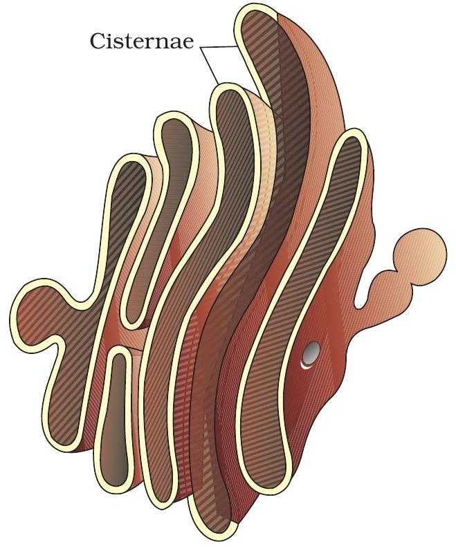
Vesicle route from Rough ER to Golgi
Protein Transit Overview
Rough ER, part of the endomembrane system, synthesises membrane and secretory proteins.
Coated vesicles bud off and move along cytoskeleton tracks to the Golgi apparatus.
The cis face receives cargo; enzymes trim, fold and tag each protein.
Finally, the trans face packages sorted proteins into vesicles for delivery or export.
Key Points:
- RER makes and folds proteins, inserts them into lumen.
- Vesicles carry cargo to Golgi cis face for modification.
- Trans face ships finished proteins — tap to confirm!