Reductionist Biology
Reductionist Approach
It views living systems through a physico-chemical lens, explaining them as interacting molecules and measurable reactions.
Why might isolating a cellular process in a test tube still leave mysteries unsolved?
Scientist Spotlight: G.N. Ramachandran (1922-2001)
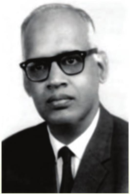
G.N. Ramachandran (1922-2001)
Trailblazer of Protein Structure
Born in Kerala, Ramachandran studied physics in Madras and Cambridge, then founded India’s first biophysics laboratory.
He created the Ramachandran Plot, mapping allowed peptide bond angles, and predicted collagen’s triple-helix model.
His ideas remain the first checkpoint for every newly solved protein structure.
Key Points:
- Invented the Ramachandran Plot (1963).
- Co-proposed collagen’s triple-helix model.
- Inspired generations of structural biologists.
What is a Cell?
Cell
Section 8.1 core idea: A cell is the smallest living unit that operates on its own. It performs metabolism, grows, and reproduces, making it the structural and functional unit of life.
Key Characteristics:
- Surrounded by a membrane enclosing cytoplasm and genetic material.
- Executes all basic life processes—metabolism, growth, response, reproduction.
- Exists alone (unicellular) or as part of multicellular organisms.
Example:
Quick check: Which instrument allowed Leeuwenhoek to first see living cells? A) Simple microscope B) Telescope
Cell Theory Timeline
Key milestones that shaped modern cell theory.
Matthias Schleiden – 1838
Studied plants; concluded every plant is composed of cells.
Theodor Schwann – 1839
Extended idea to animals; stated cells are the basic unit of all life.
Rudolf Virchow – 1855
Proclaimed “Omnis cellula e cellula” — cells arise only from pre-existing cells.
Pro Tip:
Virchow’s rule links cellular origin to life’s continuity — a key exam point.
Life Strategies
Unicellular Organisation
Multicellular Organisation
Key Similarities
Cell Shape Diversity
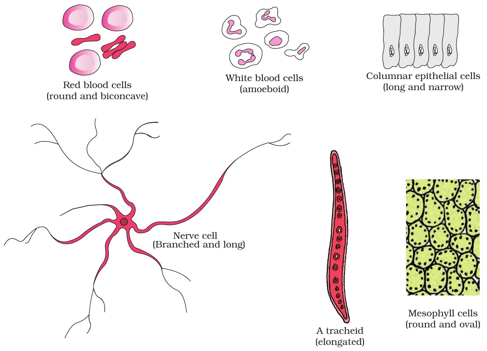
NCERT Fig 8.1: Variety of cell shapes
Form Follows Function
Figure 8.1 displays four distinct cell outlines, each shaped for its specific task.
Key Points:
- Discoid RBCs squeeze through capillaries, maximising gas exchange.
- Spindle-shaped muscle fibres shorten smoothly during contraction.
- Branching neurons span long distances to relay electrical signals.
- Amoeboid WBCs change shape to engulf pathogens.
Sizing Up Cells
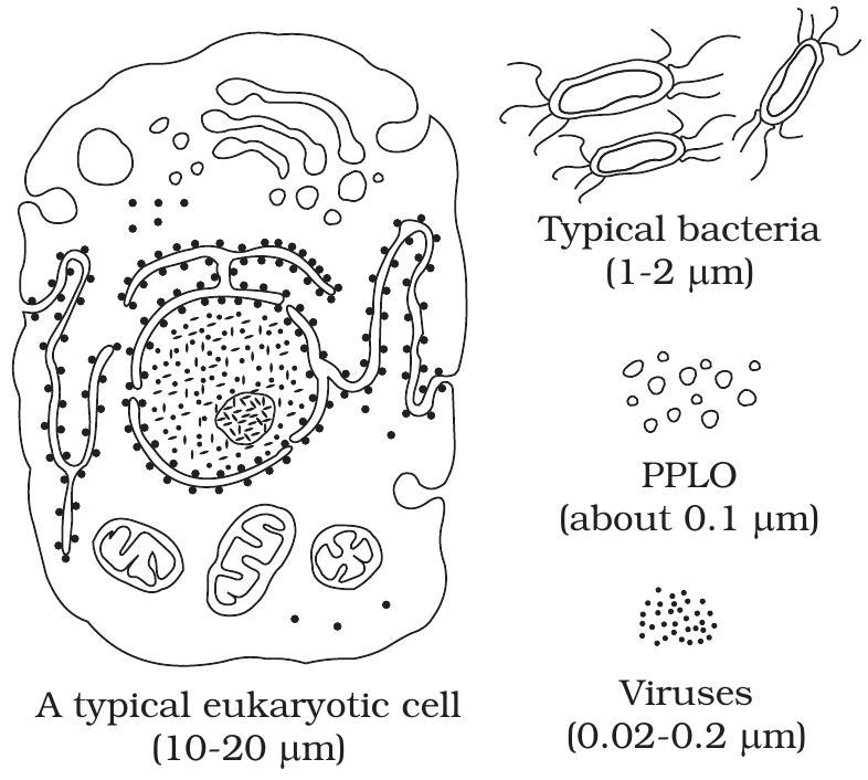
Fig 8.2 Comparison of sizes: animal cell, bacterium and virus
Cells span a huge size range
Fig 8.2 places common life forms on a shared scale.
Animal cell ≈ 20 µm, bacterium ≈ 1 µm, virus ≈ 0.1 µm.
Key Points:
- Eukaryotic cells are about 200 × wider than many viruses.
- All still rely on the same DNA, proteins and membranes.
- Micrometre (µm) suits cells; nanometre (nm) suits viral particles.
Prokaryote Primer
Prokaryotic Cell
Simplest living cells lacking a membrane-bound nucleus or organelles. Size: 1–5 µm. Reproduce swiftly by binary fission. Display three shapes—coccus (sphere), bacillus (rod) and spirillum (spiral). Bacteria and cyanobacteria exemplify them.
NCERT Section 8.4 opening: recall these hallmark traits to spot a prokaryote instantly.
Source: NCERT Biology Class 11, Chapter 8
Cell Envelope Layers
Goal: spot glycocalyx, cell wall and plasma membrane in a bacterium.
Membrane
(Capsule/Slime)
Legend:
Gram + vs Gram –
Main Points
- 1 Gram stain classifies bacteria as positive (purple) or negative (pink).
- 2 Outcome hinges on cell envelope architecture, not pigment itself.
- 3 Gram + has a thick peptidoglycan layer that locks the crystal violet-iodine complex.
- 4 Gram – features a thin wall plus an outer membrane; alcohol wash removes the dye.
Key Highlights
-
Thick peptidoglycan is an easy target for β-lactam drugs like penicillin.
-
Outer membrane of Gram – blocks many antibiotics and lysozyme.
-
Learning check: Why does penicillin work better on Gram +? — click to reveal!
Mesosomes & More
Membrane folds give prokaryotes inner “rooms” for respiration or photosynthesis.
Plasma Membrane Infolds
Bacterial plasma membrane can invaginate, creating internal sacs that enlarge surface area.
Mesosome – Respiratory Hub
Vesicular, finger-like folds hold enzymes of electron-transport chain, aid cell-wall formation and help segregate replicated DNA.
Chromatophore – Light Factory
Flattened pigment-rich sacs in cyanobacteria capture sunlight and run the light reactions of photosynthesis.
Pro Tip:
Remember: Mesosome → respiration; Chromatophore → photosynthesis. Both are plasma-membrane derivatives, not separate organelles.
Bacterial Appendages
Flagella vs Pili vs Fimbriae
These surface structures differ in shape and job: flagella drive motility, whereas pili and fimbriae enable attachment or gene transfer.
Key Characteristics:
- Flagellum – long, helical, rotating propeller; powers swimming.
- Pilus – few, hollow tubes; conjugation or specific adhesion.
- Fimbriae – numerous, short bristles; broad surface attachment.
Example:
Escherichia coli uses fimbriae to cling to intestinal epithelium before infection.
Multiple Choice Question
Question
A bacterial 70 S ribosome is made up of which two subunits?
Hint:
Unlike inclusion bodies that store reserves, a ribosome splits into one large and one small subunit.
Correct!
Well done! A 70 S ribosome is formed when a 50 S large subunit joins a 30 S small subunit.
Incorrect
Recall: 50 S + 30 S combine to give 70 S (values are not additive). Inclusion bodies are unrelated storage granules.
Eukaryotic Leap
Eukaryotic Cell
Defined by a membrane-bound nucleus, internal compartmentalisation into diverse organelles, a dynamic cytoskeleton, and linear DNA packaged with histones—upgrades that support larger, specialised life.
These features mark a clear step beyond prokaryotes and should roll off your tongue as the core eukaryotic checklist.
Plant vs Animal Cell
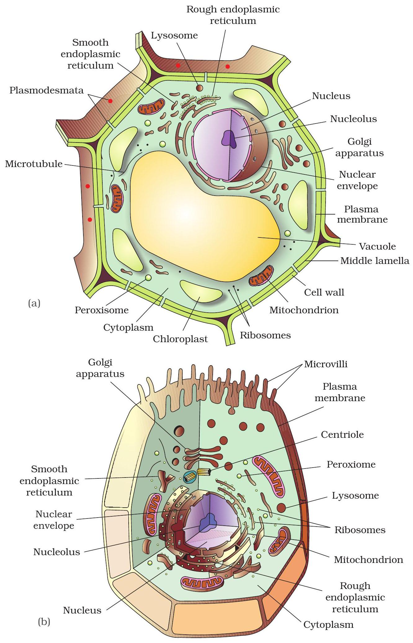
Spot the kingdom-specific organelles
Use Fig 8.3 to locate structures found only in plant cells or only in animal cells.
Recognising them lets you quickly tell the two kingdoms apart.
Key Points:
- Plant only: cell wall, chloroplasts, large central vacuole.
- Animal only: centrioles, microvilli, many small vacuoles.
Fluid Mosaic Model
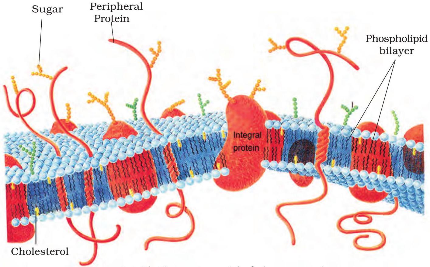
Fig 8.4: Fluid mosaic model of plasma membrane
Membrane Composition & Fluidity
Phospholipid bilayer forms a viscous, two-dimensional liquid.
Integral and peripheral proteins float within, enabling selective transport and cell signalling.
Key Points:
- Cholesterol stabilises the bilayer, moderating fluidity across temperatures.
- Lipids swap places rapidly, ensuring flexibility and self-repair.
- Proteins glide sideways, carrying molecules and relaying signals.
Transport Tactics
Main Points
- 1 Simple diffusion – solute moves from high to low concentration; no membrane protein or ATP.
- 2 Osmosis – water diffuses through a semipermeable membrane toward higher solute concentration; passive.
- 3 Active transport – carrier proteins pump molecules against the gradient using cellular ATP.
Key Highlights
-
Passive processes (diffusion & osmosis) demand zero ATP.
-
Active pumps like Na⁺/K⁺-ATPase uphold vital ion gradients.
-
Aquaporins hasten osmosis across the selectively permeable membrane.
-
Steeper concentration difference speeds every transport mechanism.
Building a Cell Wall
Trace how each plant cell wall layer forms so you can list them in order.
Primary Wall
Thin and elastic; cellulose microfibrils with hemicellulose allow the cell to expand during growth.
Secondary Wall
Deposited after growth stops; multiple cellulose layers lignified for strength. Pits keep plasmodesmata pathways open.
Middle Lamella
Outermost pectin-rich layer formed during cytokinesis; acts as a glue holding adjacent cells together.
Pro Tip:
Actual outside-to-inside order is middle lamella → primary wall → secondary wall—keep it straight in exams.
Endomembrane System
ER → Golgi → Lysosome/Vacuole
Network of membranes that moves cellular cargo. Rough ER builds proteins and lipids; Golgi tags and sorts them; vesicles deliver packages to lysosomes, vacuoles, or the plasma membrane.
Summary of Section 8.5.3, NCERT Grade 11.
Source: NCERT Biology Class XI
Endoplasmic Reticulum
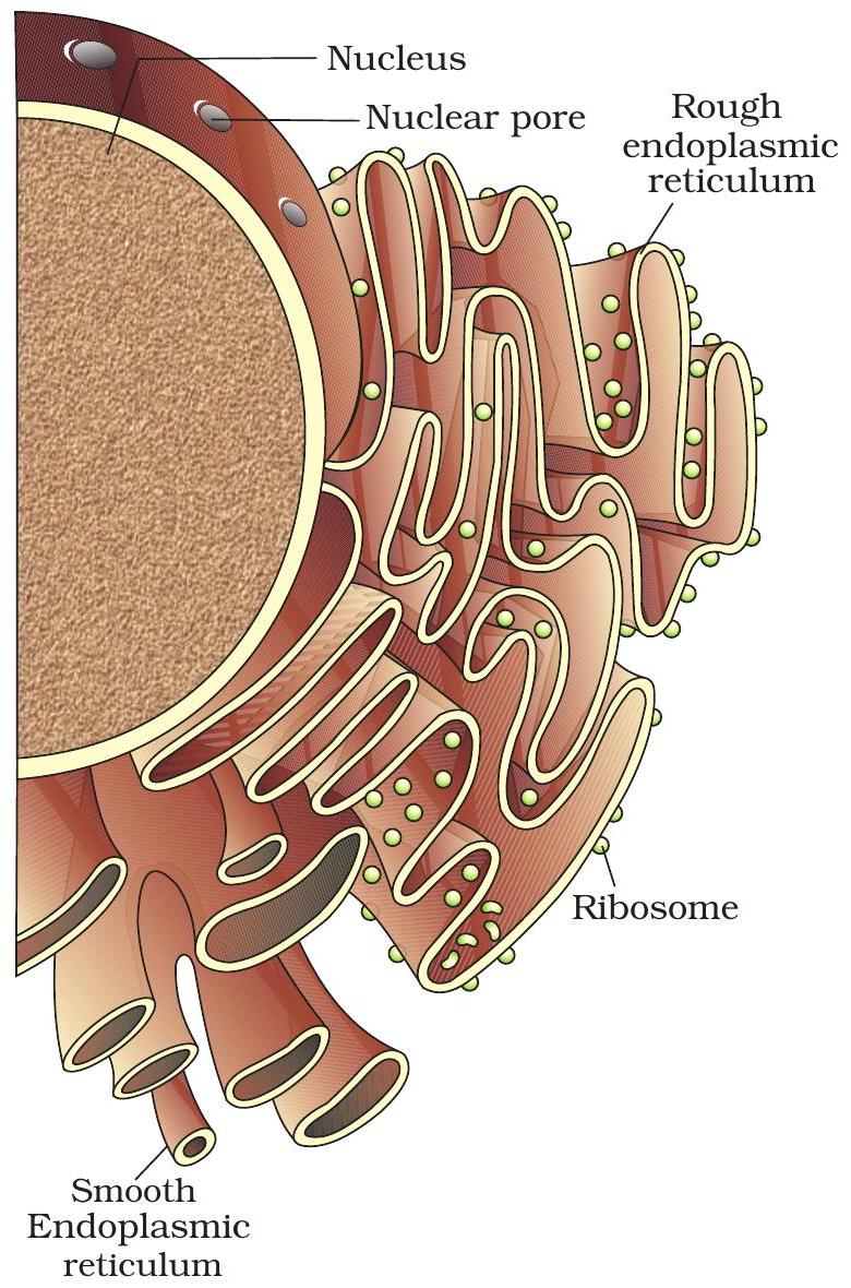
Fig 8.5 Rough and Smooth Endoplasmic Reticulum
Rough vs Smooth ER
Both form a continuous membranous network visible in Fig 8.5.
Surface features dictate their products and roles.
Key Points:
- RER: ribosome-studded cisternae.
- Synthesises and channels secretory & membrane proteins.
- SER: smooth tubules without ribosomes.
- Produces lipids, steroids & detoxifies drugs.
Golgi Apparatus
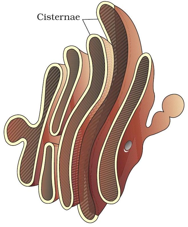
Fig 8.6 – Cis, medial and trans faces of a Golgi stack
Cis-to-Trans Maturation & Packaging
As proteins move through stacked cisternae, they are modified, tagged and finally sorted for delivery.
Key Points:
- Cis face (forming face) receives ER vesicles.
- Enzymes add sugars & phosphate tags—glycosylation.
- Cisternae mature from cis → medial → trans.
- Trans face packs cargo into vesicles for specific targets.
Lysosomes & Vacuoles
Digestive Bag vs Storage Sac
Both are single-membrane organelles; lysosomes break down materials, whereas vacuoles mainly store and maintain turgor.
Key Characteristics:
- Lysosome: acidic (pH ≈5); 40+ acid hydrolases recycle cell debris.
- Vacuole: large, water-filled; tonoplast pumps ions and stores pigments, toxins, wastes.
- Both originate from the endomembrane system and share related transport proteins.
- Core contrast: lysosome = digestion; vacuole = storage & osmotic balance.
Example:
Plant central vacuole keeps leaves firm; animal macrophage lysosome destroys engulfed bacteria.
Powerhouse Mitochondrion
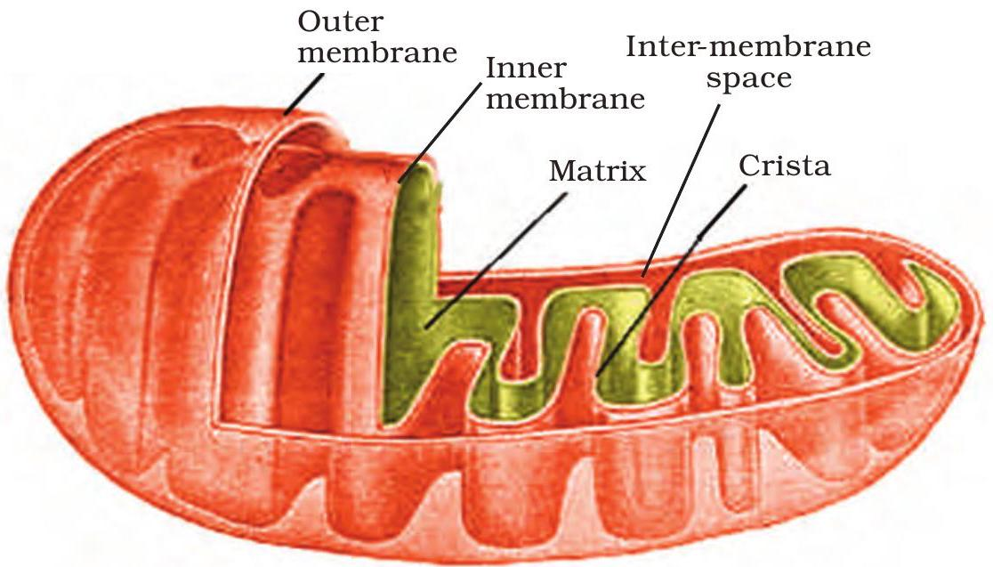
Fig 8.7 Structure of a mitochondrion
Cristae + Matrix ⇒ ATP Factory
Cristae fold the inner membrane, giving vast surface for electron transport chains that power ATP synthase.
The enzyme-rich matrix hosts the Krebs cycle, supplying NADH and FADH₂ to the cristae for oxidative phosphorylation.
Key Points:
- Double outer membrane creates inter-membrane space for proton accumulation.
- mtDNA & 70 S ribosomes allow self-replication—evidence of endosymbiotic origin.
- One glucose can yield ≈ 32 ATP within mitochondria.
Inside Chloroplasts
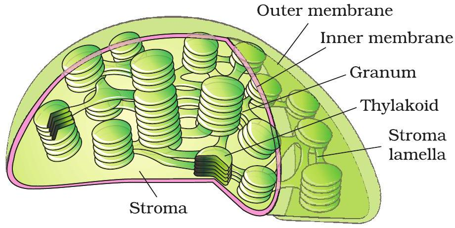
Fig 8.8 : Chloroplast ultrastructure
Locate grana, stroma & plastid DNA
Grana are coin-like stacks of thylakoids that harvest light energy.
Protein-rich stroma surrounds grana and holds Rubisco for carbon fixation.
Key Points:
- Grana → light reactions on thylakoid membranes.
- Stroma → Calvin cycle enzymes, including Rubisco.
- Circular chloroplast DNA floats in stroma with ribosomes.
Ribosome Anatomy

Fig 8.9 | Structure of an 80 S ribosome
Sedimentation Subunits
Ribosomes in cytosol are 80 S, while those inside mitochondria and chloroplasts are 70 S.
Key Points:
- 80 S ➜ 60 S large + 40 S small subunit.
- 70 S ➜ 50 S large + 30 S small subunit.
- Svedberg (S) value shows size & density; it is non-additive.
- Subunits dock together during protein synthesis, guiding mRNA.
Cytoskeleton
Dynamic Scaffold
Dynamic network of microtubules, actin and intermediate filaments that preserves cell shape, drives cytokinesis, builds the spindle and provides tracks for vesicle & organelle transport.
Microtubules = rigid tracks; actin filaments = force-generating cables. Together, they meet the functions outlined in Section 8.5.7.
Source: NCERT Biology — Section 8.5.7
Cilia & Flagella 9+2
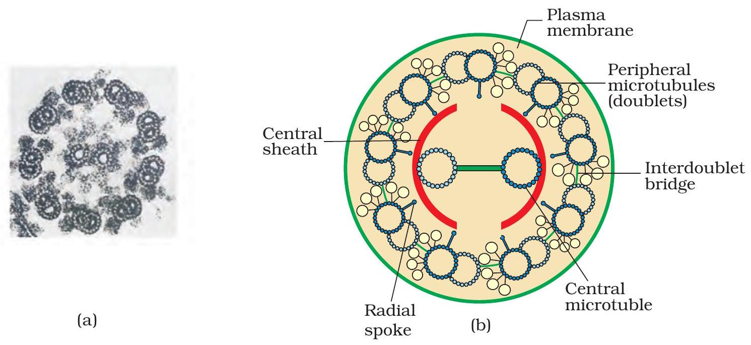
Fig 8.10 – 9+2 axoneme inside a cilium/flagellum
Axoneme: 9+2 Microtubule Core
Fig 8.10 depicts the 9 + 2 axoneme inside every cilium or flagellum.
Key Points:
- Nine peripheral doublets encircle a central pair of singlets.
- Dynein arms on A tubules walk along neighbouring B tubules.
- Radial spokes transmit signals from central pair to doublets.
- Dynein-driven sliding bends the axoneme, generating motile waves.
Centrosome Hub
Centriole
A centriole is a short cylinder of nine microtubule triplets in cartwheel pattern, forming the centrosome core that nucleates the mitotic spindle.
Section 8.5.9 — this layout explains how the spindle arises during cell division.
Nucleus Control Center
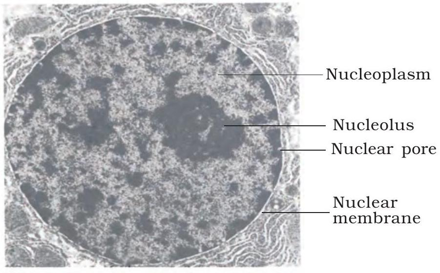
Fig 8.11 – Double-membrane nucleus with pores and nucleolus
Structure & Functions
The nuclear envelope is a double membrane that encloses chromatin and separates it from cytoplasm.
Selective nuclear pores perforate the envelope, regulating traffic of mRNA, proteins and ribosomal subunits.
Inside, the dense nucleolus synthesises rRNA and assembles ribosomal subunits.
Key Points:
- Envelope outer membrane is continuous with rough ER.
- ~100 nm nuclear pore complexes act as gated channels.
- Nucleolus disappears during mitosis and re-forms in telophase.
Chromosome Basics
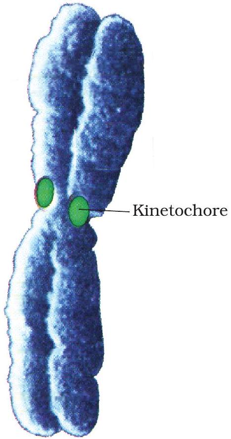
Fig 8.12 Chromatid showing centromere and kinetochore
Centromere & Kinetochore
Sister chromatids stay connected at the centromere, the chromosome’s narrow primary constriction.
A kinetochore plate assembles on the centromere and converts spindle tension into chromatid motion.
Key Points:
- Centromere glues sister chromatids together (see Fig 8.12).
- Kinetochore forms on centromere surface.
- Spindle microtubules anchor to the kinetochore.
- Motor proteins pull chromatids toward opposite poles.
Centromere Positions
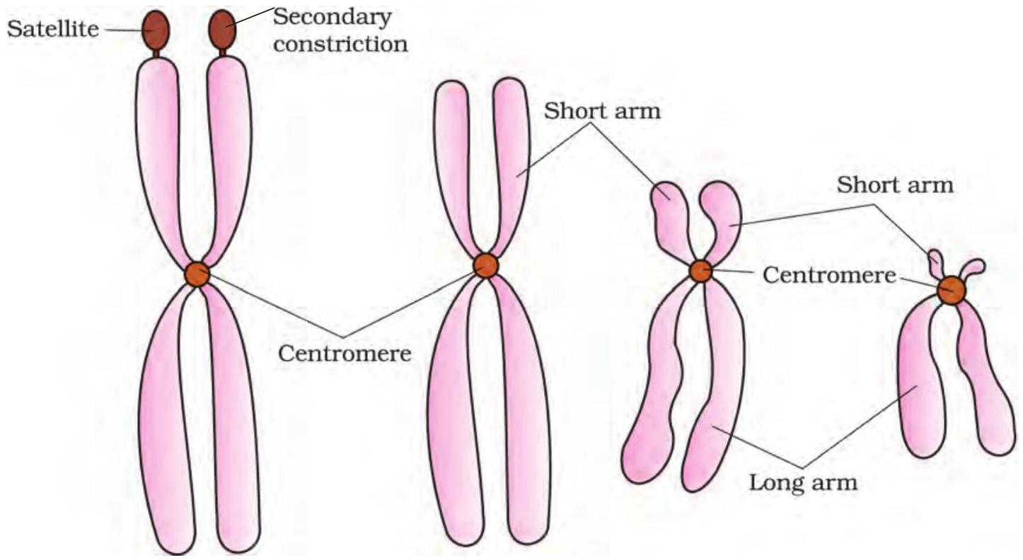
Fig 8.13 | Centromere positions and arm ratios
How arm ratio classifies chromosomes
Centromere splits a chromosome into short (p) and long (q) arms.
Shifting its position changes the p:q ratio, yielding four recognised types.
Key Points:
- Metacentric – centromere central; arms equal; forms V shape.
- Sub-metacentric – centromere slightly off; long arm exceeds short; L shape.
- Acrocentric – centromere near end; long arm dominates; J shape.
- Telocentric – centromere terminal; only one arm; I shape.