Meet the Cell Where life’s story begins at microscopic scale.

What is a Cell?

Cell

A cell is the basic structural and functional unit of life, capable of energy release, growth and self-reproduction.

Some organisms are unicellular; others are multicellular, yet each relies on its cells for structure and function. Can you name a unicellular organism?

Cell Theory

See how three scientists built the three pillars of modern cell theory.

1

Matthias Schleiden (1838)

Studied plants and concluded every plant is composed of cells.

2

Theodor Schwann (1839)

Showed animals are also cellular, making the cell the universal unit of life.

3

Rudolf Virchow (1855)

Proposed “Omnis cellula e cellula” — new cells come only from existing cells.

Pro Tip:

Remember: All organisms are cellular, the cell is life’s unit, and cells arise only from cells.

Shapes Galore

https://asset.sparkl.ac/pb/sparkl-vector-images/img_ncert/Ts2g3L11T0xHC0JGqfuxJHoFnDKuWgZL1wsWs9zi.png

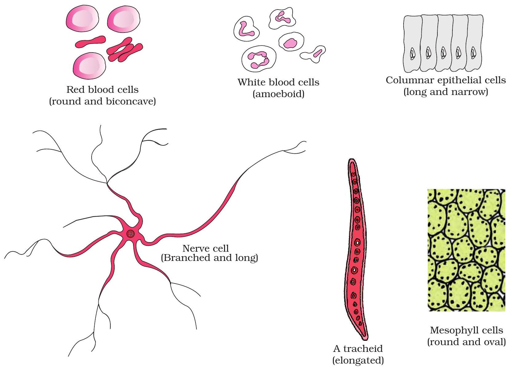
Varied cell shapes: RBC, nerve cell, epithelial

Shape mirrors duty

Cells adopt specific forms that match their roles.

Biconcave red blood cells bend easily and expose more surface for gas exchange.

A nerve cell’s long, branched axon carries impulses swiftly over distance.

Columnar or flat epithelial cells create tight sheets for protection or absorption.

Key Points:

  • RBC: biconcave disc → high surface area, easy capillary passage.
  • Nerve cell: long & branched → rapid signal conduction.
  • Epithelial cell: columnar/flat sheet → cover, protect, absorb.

Sizing Things Up

https://asset.sparkl.ac/pb/sparkl-vector-images/img_ncert/CfFVSlF9MdTVqH0uTJb8Z9H1wF6QXu9tnOfZbWCJ.png

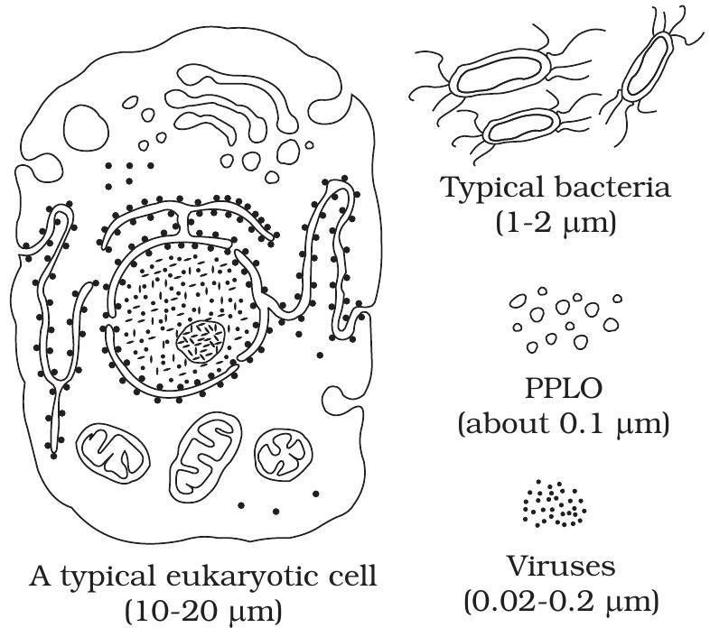
Relative Scale: Virus → PPLO → Bacterium → Human Cell

A virus measures about 20–300 nm; even the tiniest PPLO (mycoplasma) is roughly 300 nm.

Typical bacteria are 1–5 µm, whereas a eukaryotic cell often spans 20 µm or more.

Key Points:

  • Virus: 0.02–0.3 µm
  • PPLO (Mycoplasma): ~0.3 µm
  • Bacterium: 1–5 µm
  • Eukaryotic cell: 20–100 µm

Match Shape & Job

Drag each cell icon to the function whose task relies on that cell’s distinctive shape.

Draggable Items

Red Blood Cell (RBC)
Nerve Cell
White Blood Cell (WBC)
Columnar Epithelial Cell

Drop Zones

Gas Transport

Signal Transmission

Body Defense

Absorption

Tip:

Consider surface area, flexibility, or long extensions when pairing shape with function.

Prokaryote vs Eukaryote

Prokaryotic Cell

Nucleus – DNA lies in nucleoid; no nuclear membrane.
Organelles – lacks membrane-bound ones; has 70 S ribosomes only.
Size – tiny, about 0.1–5 µm.

Eukaryotic Cell

Nucleus – true nucleus with double envelope & nucleolus.
Organelles – many membrane-bound types (mitochondria, ER, Golgi).
Size – larger, usually 10–100 µm.

Key Similarities

Both have a plasma membrane enclosing cytoplasm.
DNA and ribosomes present in each cell type.
Carry out essential processes like growth & replication.

Plant & Animal Cells

https://asset.sparkl.ac/pb/sparkl-vector-images/img_ncert/8gCHuxJAIBbB0PQCBAkPPmi8jozGYkfrodGa8a8p.png

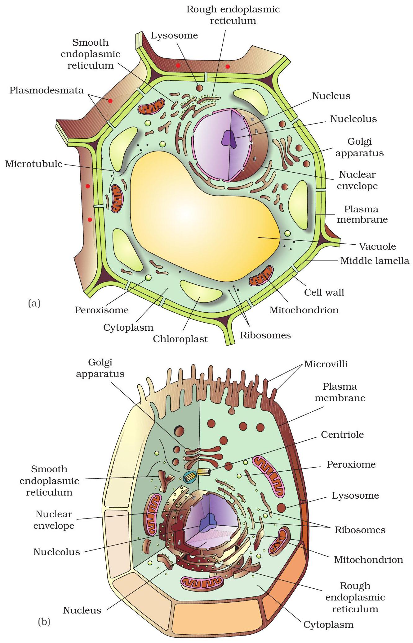
Plant vs Animal cell diagrams

Spot the unique parts

Plant and animal cells share the same core organelles for life activities.

Focus on the structures that make each cell type distinct.

Key Points:

  • Plant cell: rigid cellulose cell wall outside the membrane.
  • Plant cell: single large central vacuole for storage & turgor.
  • Animal cell: lacks cell wall, has small vacuoles and contains centrioles for cell division.
  • All other organelles are common to both cell types.

Fluid Mosaic Membrane

Fluid mosaic membrane diagram

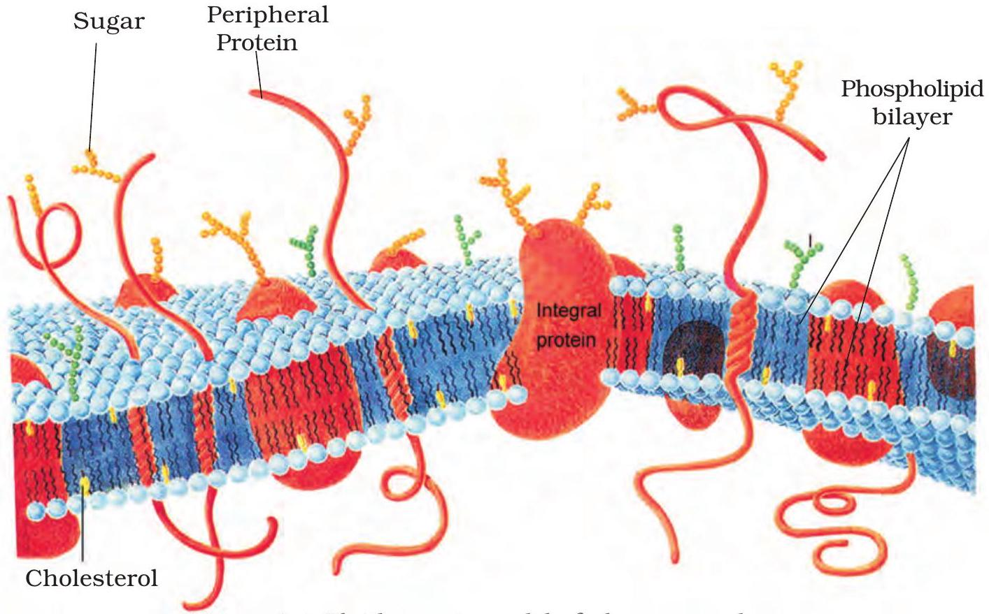
Fluid mosaic model (simplified)

A Lipid-Protein Sea

Phospholipids form a double layer; hydrophobic tails meet inside, heads face water, letting the sheet stay fluid.

Proteins float in this layer, moving laterally to relay signals and transport molecules.

Key Points:

  • Cholesterol slots between phospholipid tails, keeping fluidity steady across temperatures.
  • Membrane proteins act as channels, carriers, enzymes and receptors.
  • Lateral movement of lipids and proteins lets the plasma membrane flex and self-heal.

Endoplasmic Reticulum

https://asset.sparkl.ac/pb/sparkl-vector-images/img_ncert/9e1TluiMLQoUS2xeVc66k4ub3qkoceg0DwvZWSAl.png

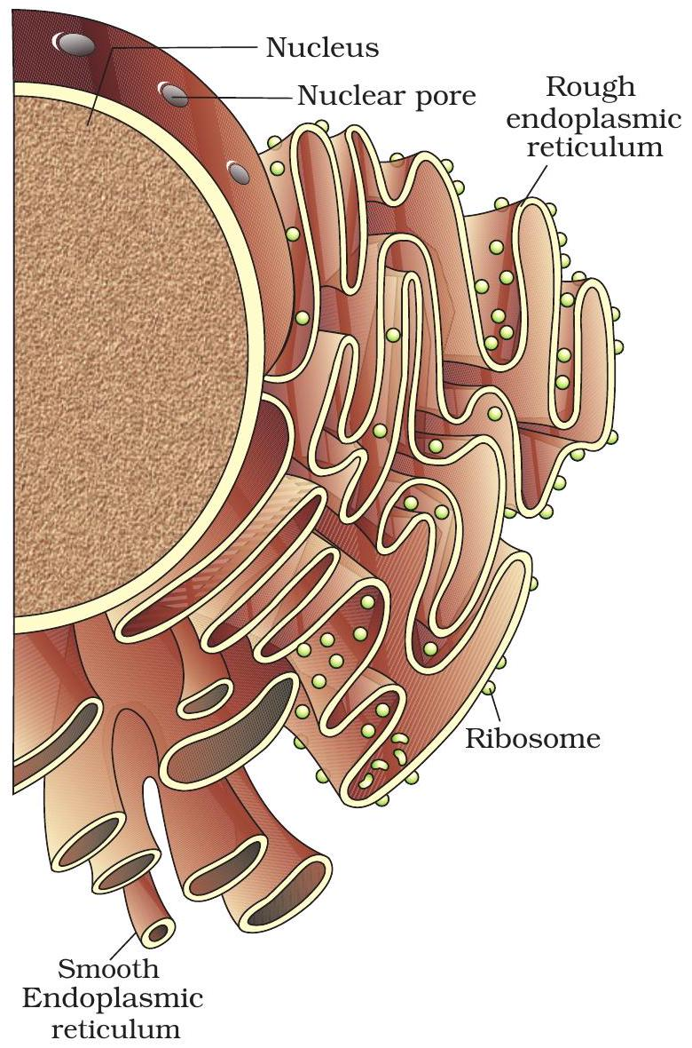
RER (ribosome dotted) and SER network inside a cell.

Rough ER vs Smooth ER

Endoplasmic reticulum (ER) forms a continuous membrane network from the nucleus into the cytoplasm.

Its rough and smooth domains look similar but perform distinct cellular jobs.

Key Points:

  • Rough ER: Ribosome-studded cisternae; synthesises secretory & membrane proteins, then sends them to the Golgi.
  • Smooth ER: Ribosome-free tubules; produces lipids, phospholipids & steroids, detoxifies drugs, stores Ca²⁺.

Golgi Packaging Hub

https://asset.sparkl.ac/pb/sparkl-vector-images/img_ncert/7UlSBEzjYFXScf6nhdzd5AmopuSkmLIZ5vewZRXN.png

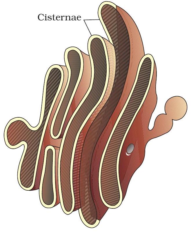
Stacked cisternae of the Golgi apparatus

Cis-to-Trans Assembly Line

Cis-face, pressed against the ER, receives fresh proteins and lipids.

Moving cisternae add or trim sugars, creating the final molecular labels.

Trans-face packs the labeled cargo into vesicles that head to membranes or outside.

Key Points:

  • Cis-face = receiving dock
  • Trans-face = shipping desk
  • Vesicles = delivery trucks

Mitochondrion

Mitochondrion

Double-membrane organelle that makes ATP, earning the name “power house” of the cell.

Key Characteristics:

  • Cristae – inner membrane folds that boost enzyme-loaded surface area.
  • Matrix – central fluid containing DNA, ribosomes and Krebs-cycle enzymes.
  • ATP output – oxidative phosphorylation here supplies most cellular energy.

Chloroplast Factory

Chloroplast diagram highlighting grana and stroma

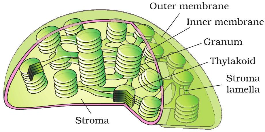
Grana stacks & stroma in a chloroplast

Grana & Stroma Roles

Light reaction happens on grana—flattened thylakoid stacks.

Chlorophyll here captures sunlight to form \( \text{ATP} \) and \( \text{NADPH} \).

Dark reaction (Calvin cycle) unfolds in the surrounding stroma.

Stroma enzymes use CO₂ plus the ATP & NADPH to build glucose.

Key Points:

  • Grana = site of light reaction, energy capture.
  • Stroma = site of dark reaction, carbon fixation.
  • Structure keeps the two phases organised and efficient.

Ribosome

Ribosome

Universal molecular factory: two subunits clamp mRNA and catalyse peptide-bond formation, converting codons into a growing polypeptide during translation.

Key Characteristics:

  • 70S (50S + 30S) in prokaryotes & organelles; 80S (60S + 40S) in eukaryotic cytoplasm.
  • Small subunit reads mRNA; large subunit forms peptide bonds via rRNA peptidyl-transferase.
  • Multiple ribosomes can form a polyribosome for rapid protein assembly.

Example:

Bacterial 70S ribosomes assemble enzymes; human 80S ribosomes synthesise haemoglobin.

Key Takeaways

Cell theory: all life is cellular, and cells arise only from existing cells.

Prokaryotes are small and simple; eukaryotes are larger and compartmentalised.

Organelles are specialised workstations—mitochondria make ATP, ER and Golgi process molecules.

Fluid plasma membrane regulates material exchange and enables cell-to-cell communication.

Next Steps

Practise labeling organelles on diagrams and link each to its specific function.

Thank You!

We hope you found this lesson informative and engaging.