Shapes Tell Stories
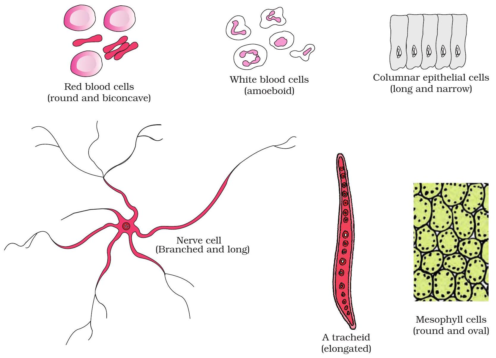
Neuron & RBC illustrate how shape serves function.
Form Mirrors Function
Cell shapes evolve to match specific tasks, linking structure with performance.
Key Points:
- Neuron: Long, branched shape carries signals swiftly across the body.
- RBC: Thin biconcave disc increases surface area for rapid oxygen exchange.
- Muscle fibre: Cylindrical length lets contractile proteins slide for movement.
- Guard cell: Kidney shape opens or closes stomata to regulate gas flow.
Sizing Up Cells
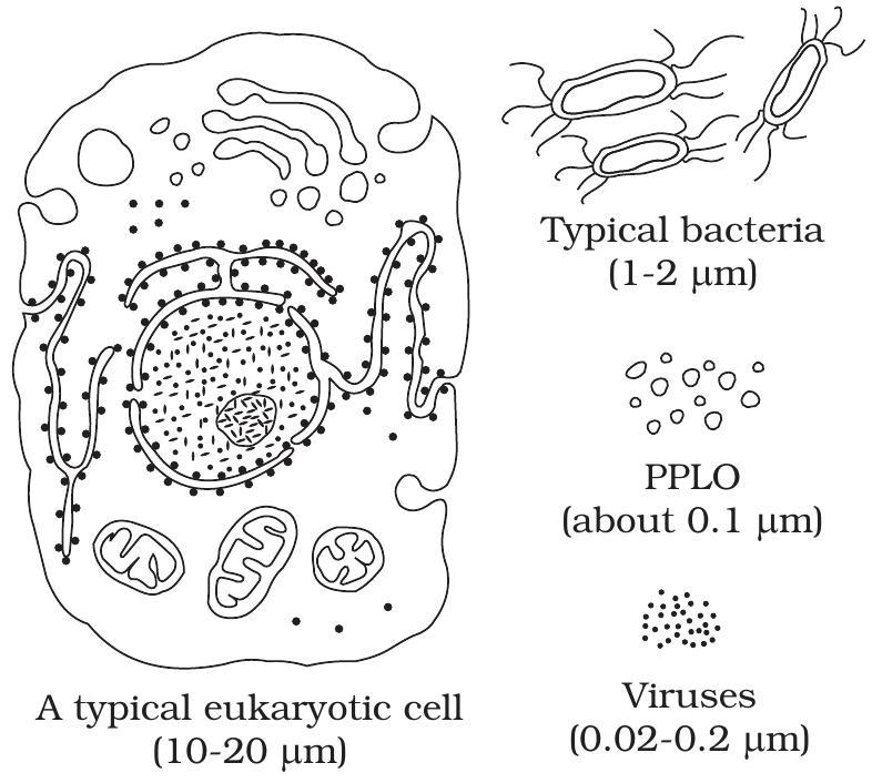
Relative sizes (not to scale)
Cell dimensions in μm
1 μm (micrometre) = 10⁻⁶ m; this unit sets the scale for cell biology.
Comparing sizes helps explain how surface area limits functions like nutrient uptake.
Key Points:
- Viruses: 0.02 – 0.3 μm, visible only with an electron microscope.
- Bacteria: 1 – 5 μm; typical prokaryotic size, light-microscope range.
- Eukaryotic cells: 10 – 20 μm; some specialised cells reach 100 μm.
Fluid Mosaic Membrane
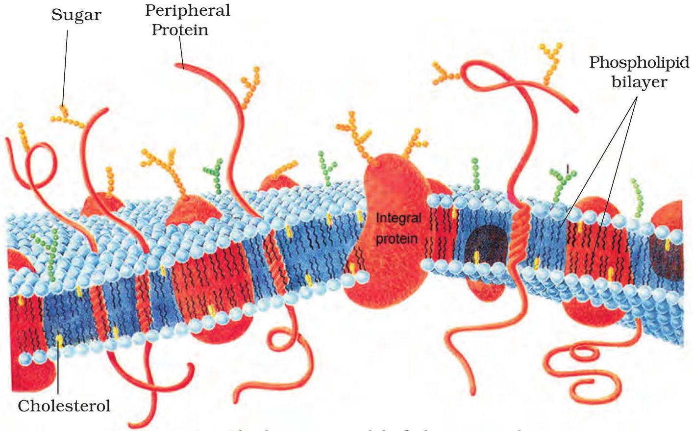
Fluid mosaic model (Singer & Nicolson, 1972)
Lipids drift, proteins skate
The plasma membrane is a fluid phospholipid sea that heals and flows.
Proteins move within this lipid matrix, creating the ever-changing mosaic.
Key Points:
- Phospholipids + cholesterol give flexibility and selective permeability.
- Proteins drift laterally but rarely flip between leaflets.
- Dynamic membrane explains cell growth, endocytosis, and self-repair.
Label the Membrane
Drag each label onto the correct feature of the plasma membrane diagram to show you can identify its components.
Draggable Items
Drop Zones
Phospholipid head
Hydrophobic tail
Integral protein
Peripheral protein
Cholesterol
Tip:
Remember: hydrophilic heads face water; hydrophobic tails hide inside the bilayer.
SA:V — The Maths
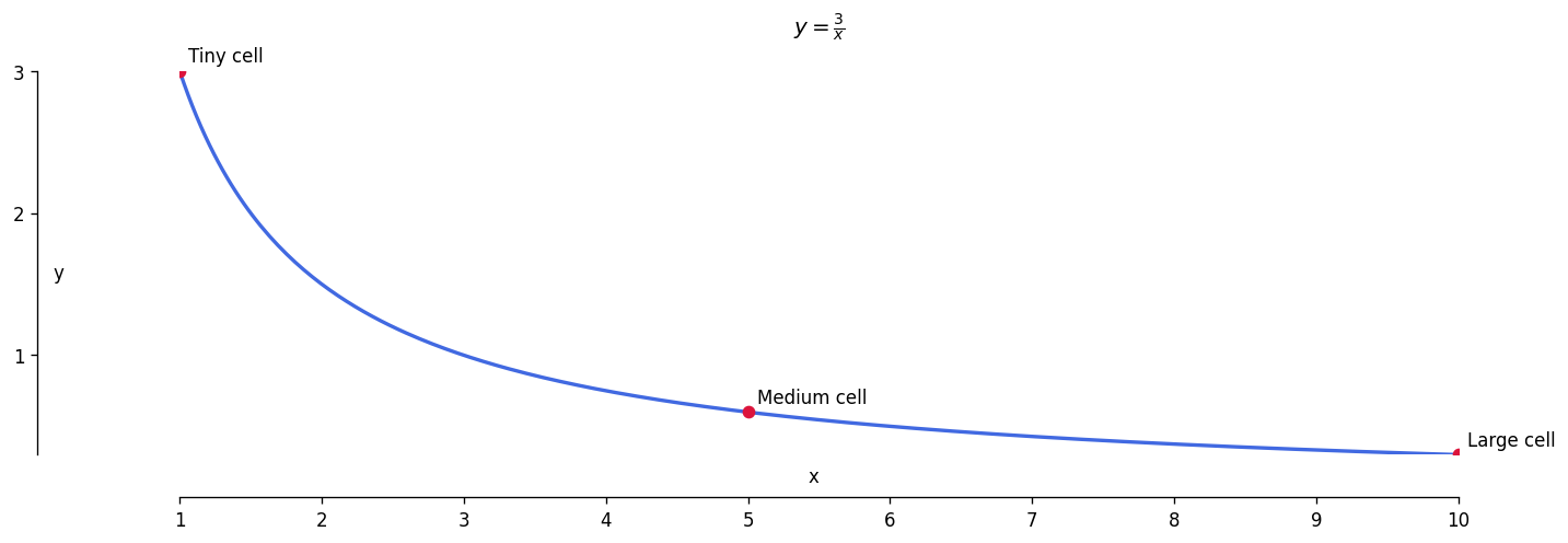
Surface area-to-volume ratio vs cell radius
Interpreting the Curve
For a sphere, \( \text{SA:V} = \frac{3}{r} \). Doubling radius halves the ratio.
The graph’s steep inverse drop shows how a slight size increase quickly lowers available surface for exchange.
Key Points:
- Inverse hyperbola: slope drops fastest at small radii.
- Lower SA:V limits diffusion-based nutrition and waste removal.
- Cells divide to regain a higher surface area-to-volume ratio.